


Description
This equipment is part of Surface Characterisation of the Multidisciplinary Characterisation Facility.
The technique involves illuminating a sample with a monochromatic light source, such as a laser. Light that is scattered has a different colour to the light source and can be analysed using a highly sensitive spectrometer. Coupling of a confocal microscope to a Raman spectrometer enables surfaces to be mapped with high spatial resolution, giving an image of the uniformity (homogeneity) of the material.
Uses / Applications
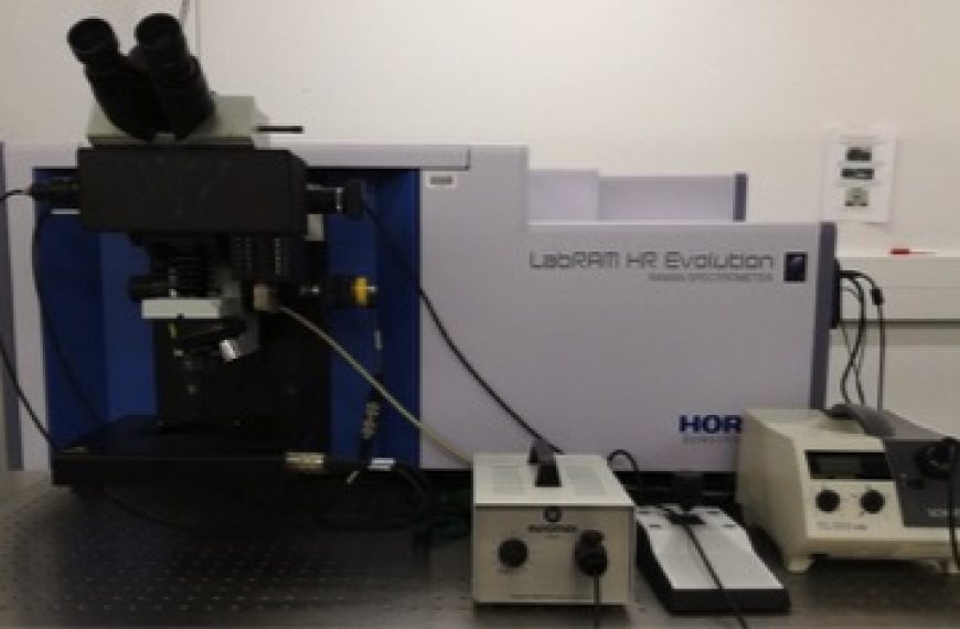
The Horiba LabRAM Evolution HR Raman Spectrometer is a fully integrated confocal Raman microscope instrument with three laser sources (325nm, 488nm and 633nm), two diffraction gratings (600g/mm and 1800g/mm), motorised XYZ stage and free-space microscope. The instrument is controlled using LabSpec 6 software.
Specification
Laser Wavelength and Power: 325nm – 20mW, 488nm – 100mW (adjustable), 633nm – 17mW.
Diffraction Grating: 600g/mm and 1800g/mm
Laser (Rayleigh) Filtering Type: Edge
Microscope: Olympus BXFM Free-Space Microscope with trinocular head
Microscope Objectives: VISIBLE – 5X (NA 0.10), 10X (NA 0.25), 20X (NA 0.4), 50X SLWD (NA 0.35), and 100X (NA 0.90). UVB – 15X (NA 0.32) and 40X (NA 0.50).
Polarisation Options: Half-Wave Plate (VIS and NUV) for incident light and Analyzer (VIS and UV) for scattered light.
Spectral range: 220-1100nm (upgradable to 2200nm)
CCD: Open Electrode CCD air-cooled to -60°C
Märzhäuser mapping stage specification: Resolution XY = 100nm, Z = 10nm. Maximum range X = 75 mm – Y = 50 mm.
Duoscan mapping resolution: XY = 50nm
Rapid mapping mode: Swift ultra-fast mapping (max scan time per point = 1s)
Software: LabSpec 6