


Description
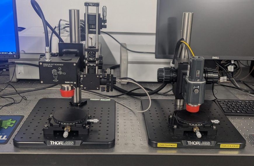
The Ganymede Series systems utilise spectral-domain optical coherence tomography (SD-OCT) technology, which is a method which provides high-resolution, cross-sectional imaging based on low-coherence interferometry. In SD-OCT, a swept source laser is incident on the sample, from which the backscattered light from different depths is analysed using a detector which enables high-resolution imaging of internal structures in biological tissues and materials.
The system is optimised primarily for high-speed imaging with A-scan rates that reach up to 248 kHz. In OCT, an A-scan represents a single depth profile, while B-scans are formed by stacking multiple A-scans together to create cross-sectional (2D) images. With higher A-scan rates, the system can acquire images faster.
ThorImageOCT software provides real-time image acquisition, processing, and data visualisation. The software includes advanced features such as:
-
Live Display & Averaging which aids in reducing measurement noise and therefore enhances image quality.
-
3D Volume Rendering which enables the user to dissect volumetric datasets interactively.
-
Time-Series Imaging allows the operator to capture dynamic changes over time.
Uses / Applications
Biomedical Research: Tissue imaging, collagen analysis, vascular studies, and monitoring biological processes.
Material Science: Subsurface defect detection, structural analysis, and quality control.
Industrial Inspection: Non-destructive testing of layered structures and product integrity verification
Specification
-
Micron-scale resolution for detailed cross-sectional and volumetric imaging.
-
High-Speed Imaging allows for A-scan rates of up to 248 kHz for real-time imaging of moving samples. Doppler and Speckle Variance Imaging allowing visualisation of vascular structures and various biological processes.
-
900 nm centre wavelength, 3.0 µm Resolution in air, 5.5 to 248 kHz A-scan/Line Rate, 1.9 mm (air)/ 1.4 mm (water) imaging depth, 3.0µm/2.2 µm axial resolution