Description
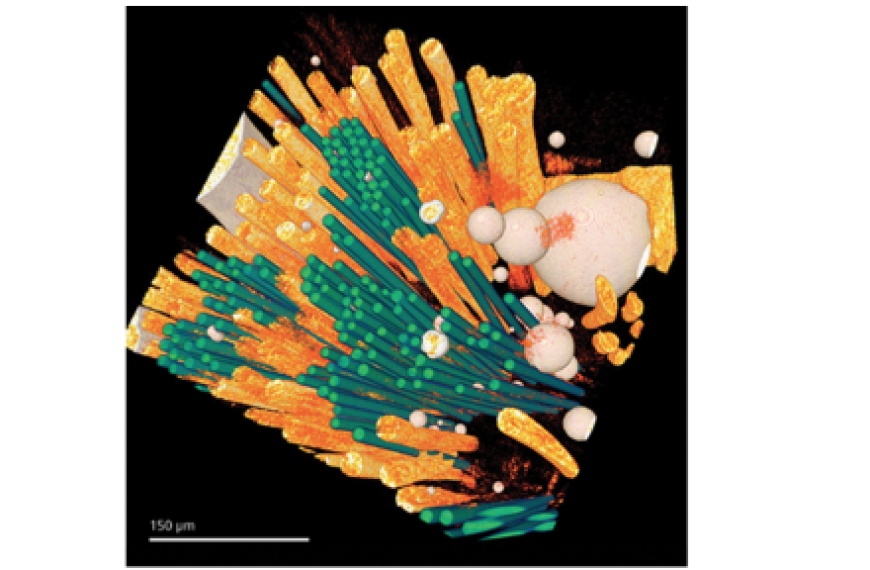
The 3D and 4D μm-scale x-ray computed tomography (XRCT) microscope is used for in-situ characterisation of the composition, deformation and damage development of materials at length scales on the order of 1 µm. It is useful for determining the relationship between processing and microstructure, for observing fracture mechanisms, investigating properties at multiple length scales, and for quantifying and characterising microstructural evolution.
Uses / Applications
Previous projects have included the characterisation of indentation of battery cathode materials, the observation and analysis of the porosity of 3D printed ceramics and time dependant observation of diffusion from medical implant devices.


