This website uses cookies so that we can provide you with the best user experience possible. Cookie information is stored in your browser and performs functions such as recognising you when you return to our website and helping our team to understand which sections of the website you find most interesting and useful.
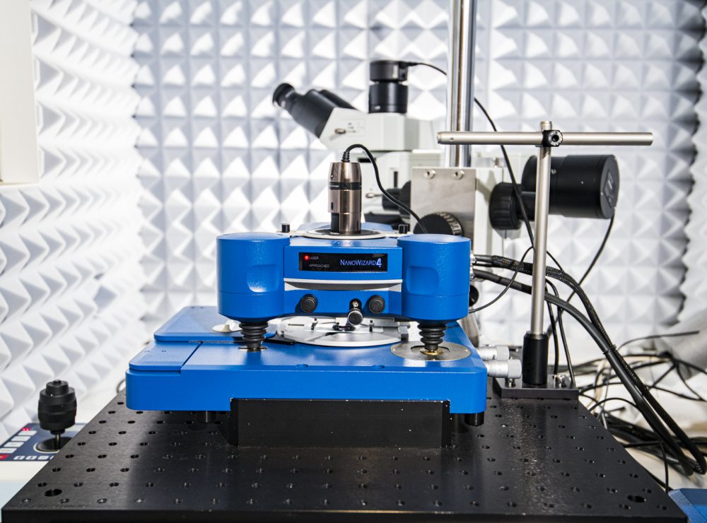
Atomic Force Microscope (AFM) with in-situ analysis
Atomic force microscope (AFM) for investigating interactions across a surface, with specialist modes for 2D materials
- Partner:The University of Manchester
- Facility:National Graphene Institute (NGI)
Or call us now on 0161 275 8382
Detailed Description
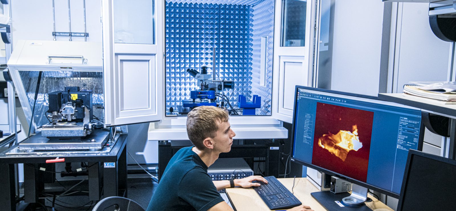
The atomic force microscope (AFM) is a powerful tool for investigating interactions across a surface.
It can create images of the surface morphology, measure the friction properties, or determine the conductivity across a sample rapidly with nanometre resolution. These modes can be used to investigate graphene/polymer composites to design improved materials. It can also analyse samples in a liquid environment and at a range of different temperatures, as well as perform electrochemical measurements.
Uses/Applications
Users can look at how supercapacitors or batteries made from 2D materials transform as they are charged and discharged, allowing for discovery of where the structure starts to fail. This will allow for the design of longer lasting and more efficient batteries.
Control
- Closed loop XY scanner
- XY scan range capable of at least 100 um x 100 um
- XY resolution <0.5 nm [Define how this is measured]
- Z range > 10 um (Z resolution <0.05 nm with acoustic and vibration isolation)
- Cantilevers must be easy to exchange (plug-in, plug out type; exchanges in minutes; no technical expertise required)
Sample stage
- Low drift
- Automated course stage control over large range (>10 mm)
- Ability to house large samples (>50 mm XY diameter, >10 mm thickness)
- Easy access for quick sample changes (exchange in minutes) and in-situ measurements
Imaging
- Multiple imaging modes including at least: contact mode (height, deflection), non-contact modes (height, phase), lateral force mode, conductive AFM, nano-indentation and nano-lithography
- Force curve mapping (for mechanical properties)
- High resolution imaging in a liquid environment
Optics
- At least 20x objective diffraction limited, long working distance >15 mm
- On axis vision of sample surface and cantilever
- High quality CCD (>4 MP)
- Large field of view (>500 um diameter)
- Adjustable intensity of light source
Electrochemical imaging
- Electrochemical cell with high chemical stability; Stable for aqueous and organic solvents, as well as acids and bases; 2 Teflon, 2 peek, 2 fused silica supplied
- Selectable area for analysis for EC cell
- Multiple ports for easily interchangeable electrodes
- Compatible with multiple potentiostats (already owned)